
Imagine the nightmare scenario of your loving pet of 10 years suddenly turning on you. This horror played out in real life for Jacxson’s owner. The previously happy Havanese had become increasingly aggressive toward his mom, and it was all because of a brain tumor the size of a kidney bean.
Jacxson’s tumor, a meningioma, was located at his frontal lobe, an area of the brain responsible for regulating behavior and personality. The small mass transformed Jacxson from a sweet, loving pup into a terror.
Meningiomas are the most common brain tumors of dogs and cats. This type of tumor typically develops on the meninges – protective layers that envelop the brain. As they grow, these tumors press onto the brain and can cause a variety of neurologic abnormalities including seizures, blindness, and personality changes. Additionally, meningiomas often incite an inflammatory response in the surrounding brain
tissue.
After a visit and series of tests with board-certified veterinary neurologist and neurosurgeon Dr. Jill Narak, Jacxson was diagnosed with this type of bean-sized tumor and deemed a good candidate for surgery. Luckily his “mean bean” had not yet caused seizures or blindness and Dr. Narak was able to remove the tumor in its entirety.
Once removed, Jacxson’s tumor was graded as the least aggressive form of meningioma (Grade I). This was wonderful news for Jacxson because it meant he was essentially cured and would not require chemotherapy or radiation therapy, as is oftentimes necessary after a cancer diagnosis.
Now that Jacxson is free of his “mean bean,” his owner reports that he is 90% his normal self and a nice dog again! Although brain surgery is a scary proposition for many owners, Jacxson’s case illustrates the place for invasive surgery with curative intent. Dr. Narak at Veterinary Referral Surgical Practice in Roswell, GA is happy to discuss when such interventions might be beneficial for your pet.

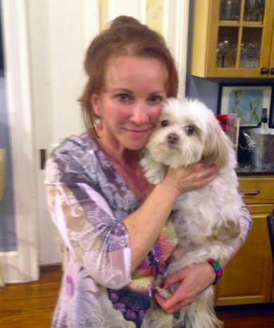
Jacxson, a 10 yr old Havanese afflicted by an intracranial meningioma.

A magnetic resonance image (MRI) shows Jacxson’s tumor located in his right frontal lobe.

Jacxson’s surgery. His nose is to the right. His skin, skull, and dura mater (outer layer of meninges) have been incised, removed, and reflected. The brain and associated blood vessels can be seen, as well as the dark red tumor at the bottom right portion of the skull opening.

Jacxson’s tumor. The meningioma, or “mean bean,” was removed in its entirety and submitted to a laboratory for analysis.

Jacxson recovering smoothly from his brain surgery.

Jacxson’s meningioma (Grade I) was deemed not to be malignant after pathologic analysis.